
A lump appears behind the knee, sometimes without pain, sometimes with discomfort during bending. This localized swelling often corresponds to a popliteal cyst, also known as a Baker’s cyst. It forms when synovial fluid accumulates in a pocket at the back of the joint. Understanding its causes and recognizing warning signs can help avoid unnecessary complications.
Popliteal cyst and intra-articular injury: the link revealed by imaging
In adults, a Baker’s cyst is rarely an isolated anomaly. In the vast majority of cases, the cyst indicates an underlying intra-articular injury: meniscal damage, osteoarthritis, synovitis, or chronic knee effusion. The excess joint fluid migrates through a small natural passage (a capsular hiatus) to the popliteal fossa, where it forms the cystic pocket.
This is why treating only the cyst without exploring the knee is akin to emptying a bucket without turning off the tap. When a doctor discovers a popliteal cyst, they systematically look for the joint cause that fuels the production of synovial fluid.
To better understand the situations that warrant a prompt consultation, you can read the article from Bien et Vous that details the vigilance criteria.
Baker’s cyst symptoms: from mild discomfort to sharp pain
Have you noticed a tension behind the knee while squatting? This is one of the first signs. The popliteal cyst manifests very variably depending on its size and condition.
When the cyst remains silent
Many popliteal cysts cause no symptoms. They are discovered by chance during an MRI prescribed for knee pain of another origin. A cyst visible on imaging is not necessarily responsible for the pain. This distinction is crucial to avoid unnecessary treatments.
When discomfort sets in
A growing cyst can lead to:
- A sensation of tension or stiffness at the back of the knee, especially in full flexion (squatting position, climbing stairs)
- A visible and palpable swelling in the popliteal fossa, with volume that can vary from day to day
- A progressive limitation of joint range of motion when the cystic pocket compresses nearby structures
When the cyst ruptures
This is the most misleading situation. A ruptured popliteal cyst releases its liquid content into the calf. The result: sudden calf pain with swelling, redness, and warmth. This presentation closely resembles deep vein thrombosis (phlebitis). This is referred to as pseudothrombophlebitis.
Without imaging, it is impossible to distinguish between the two. A prompt consultation is then necessary, not because the rupture of the cyst is serious in itself, but because a true venous thrombosis must be urgently excluded.
Common causes of a knee cyst
The popliteal cyst does not appear without reason. It indicates an excess of joint fluid, which is itself related to irritation or injury in the knee. Three mechanisms dominate in adults.
Knee osteoarthritis remains the most common cause after the age of fifty. Worn cartilage causes chronic inflammation of the synovial membrane, which produces more fluid. This surplus migrates to the popliteal fossa.
Meniscal injuries (tear, flap) disrupt joint mechanics and maintain an effusion. On an MRI, it is not uncommon to see a popliteal cyst associated with a torn meniscus, with the cyst being the consequence and not the cause of the pain.
Inflammatory synovitis (rheumatoid arthritis, gout) can also trigger the formation of a cyst by significantly increasing synovial fluid production.

Diagnosis of the popliteal cyst: ultrasound or MRI
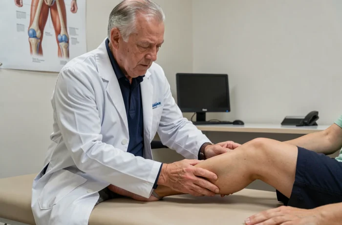
The doctor begins with a clinical examination. A soft, fluctuating mass located in the popliteal fossa quickly points to a Baker’s cyst. The next step is imaging, and ultrasound is the first-line examination.
It is accessible, non-invasive, and cost-effective. It visualizes the cyst, assesses its volume, detects any septations, and checks if the content is purely liquid. In Doppler mode, it also helps rule out deep vein thrombosis, which is particularly useful in cases of calf pain.
The MRI is used in two specific situations:
- When the doctor suspects an associated meniscal, cartilaginous, or ligamentous injury that requires a complete assessment of the knee
- When the popliteal mass has an atypical appearance (solid component, heterogeneous morphology, infiltrative behavior) that deviates from the classic presentation of a Baker’s cyst and warrants excluding another pathology
Standard X-rays have a limited role. They do not show the cyst itself but provide an overview of the bony condition and any potential presence of osteoarthritis.
When to consult a doctor for a knee cyst
A painless, stable popliteal cyst discovered by chance does not require immediate treatment. However, certain signals necessitate a consultation without delay.
Consult quickly if the calf suddenly swells, becomes painful, warm, or red. This presentation may correspond to a rupture of the cyst or a venous thrombosis, and only imaging can clarify the situation. Delaying the diagnosis of phlebitis exposes one to serious complications.
A cyst that regularly increases in volume or limits knee flexion in daily life also deserves medical advice. The doctor will investigate the underlying joint pathology (osteoarthritis, meniscal injury) and adjust treatment to the actual cause.
Finally, any popliteal mass whose appearance changes (becoming hard, irregular, or painful at rest) should prompt a reconsideration of the diagnosis. An atypical mass behind the knee is not always a Baker’s cyst, and additional imaging may be necessary to rule out other pathologies.
The popliteal cyst remains, in most cases, a benign consequence of a treatable joint problem. Correctly identifying it, understanding what fuels it, and monitoring for signs of rupture or atypia allows for timely intervention, neither too early nor too late.